Practical activities bio year 9
Living beings are classified in 5 groups (Kingdoms):
Animals, Plants, Fungi, Protoctists (protozoos + algae) [All EUKARYOTAE] and
Monera (Bacterias) [prokaryotae].
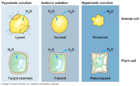
 2A) The membrane is composed of phospho-lipids and proteins.
 2B) The membrane is a boundary, selective barrier & protects the cell.
The cytoplasm contains: organelles, hyaloplasm, cytoskeleton and inclusions.
4) Ribosomes make proteins. They get the "recipe" in a RNA molecule (copy of a small region of DNA from the nucleus).
Animal cell vs Plant cell .
OUT of the mitochondria, withOUT Oxygen, in the cytoplasm, cells can break down glucose in --> 2 molecules of lactic acid and get 2ATP, or --> 2 alcohol + 2 CO2 + 2ATP
9) Chloroplast: 💥(only in plants and algae, and some protozoos).

--> Types of tissues in animals (IMG): Atlas Michigan
--> Systems in the human body. IMG


T4 Digestion 💥
Digestion is the process of breaking down food into smaller molecules that can be absorbed by the body. It may be mechanical or chemical.
It begins in the mouth with mechanical digestion or mastication, grinding food into smaller pieces with saliva. Chemical digestion begins in the mouth with the action of enzymes like the salivary amylase, which breaks down starch into disaccharides (maltose). The salive also contains lysozyme that kills bacteria and mucin to lubricate the bolus.

From the stomach, the chyme moves to the duodenum (first part of the small intestine), where it is further broken down by enzymes from the pancreas, liver (gall or bile), and small intestine itself. Here the pH increases to 8, to inactivate the gastric enzymes and activate the new ones. Pancreatic enzymes break down carbohydrates, proteins, and lipids. Bile from the liver, which is stored in the gallbladder, has lipase that breaks down lipids. And the chyme becomes the chyle.
The chyle (chyme + bile + pancreatic juices) contains carbohydrates broken down into monosaccharides, proteins broken down into amino acids and lipids broken down into fatty acids and glycerol. The DIGESTION is done.

The ABSORPTION: That mixture will move on to the 2nd and 3rd part of the small intestine: jejunum and ileum, where most of the nutrients are absorbed into the bloodstream and then transported to the liver. (The liver will release nutrients slowly along the rest of the day, as they are needed).
The rest continues into the large intestine or colon, which is colonised by bacteria (intestinal flora). They protect us, ensure the mineral balance for the water absorption, and release vitamin K (for coagulation) and B. The waste products will eventually be excreted from the body as faeces, through the rectus and anus. Bacteria also produce gases (methane and others).
Key terms:
Enzyme: A protein that speeds up a chemical reaction.
Intestine: A long, coiled tube that is part of the digestive system.
Nutrient: A substance that provides energy, builds and repairs tissues, or regulates body functions.
Saliva: A fluid produced in the mouth. It has lysozyme to kill bacteria, mucin to lubricate and amylase.
Small intestine: The longest part of the intestine. Duodenum (digest) + Jejunum + Illeum (absorpt).
Large intestine: The last part of the intestine: ascendent colon, transverse colon and descending colon.
T5 Respiratory system
The Respiratory System: A Journey of Gas Exchange

The respiratory system, a remarkable network of intricate structures, is responsible for the crucial process of gas exchange, ensuring the continuous supply of oxygen to every cell in our bodies and the removal of carbon dioxide, a waste product of cellular respiration. This elaborate system, composed of the respiratory airways, lungs, and muscles, orchestrates the movement of air into and out of the body, enabling the exchange of these life-sustaining gases.
The Respiratory Airways: A Pathway for Air Intake: The journey of air into our bodies begins with the nostrils, the external openings that act as gateways for inhaled air. The air then passes through the nasal passages, where it is warmed, moistened, and filtered, removing dust particles and other impurities. Next, the air enters the pharynx, a common passageway for both air and food. From here, it moves into the larynx, commonly known as the voice box, where vocal cords vibrate to produce sounds.
Following the larynx, the air enters the trachea, a rigid tube supported by cartilaginous rings, which help keep it open during inhalation. The trachea branches into the right and left bronchi, which further divide into smaller bronchioles, eventually terminating in microscopic air sacs called alveoli.

The Movements of Intercostal Muscles and Diaphragm: A Symphony of Breath: The process of breathing, the rhythmic movement of air into and out of the lungs, is orchestrated by a coordinated interplay of muscles. The intercostal muscles, located between the ribs, play a crucial role in expanding and contracting the chest cavity. As the intercostal muscles contract, they raise the ribs, increasing the volume of the chest cavity, thereby creating a partial vacuum that draws air into the lungs during inhalation.
In contrast, during exhalation, the intercostal muscles relax, allowing the ribs to fall back down. This decrease in chest cavity volume pushes air out of the lungs. Simultaneously, the diaphragm, a dome-shaped muscle at the base of the lungs, also plays a vital role in breathing. The diaphragm contracts during inhalation, flattening and expanding the chest cavity further, aiding in the intake of air.
Gas Exchange: The Oxygen and Carbon Dioxide Exchange at the Alveoli: The alveoli, the tiny air sacs at the end of the bronchioles, serve as the primary sites for gas exchange. Surrounded by a network of capillaries, the smallest blood vessels in the body, the alveoli provide an ideal interface for the diffusion of gases.
As oxygen-rich air enters the alveoli during inhalation, it diffuses across the thin walls of the alveoli and the capillaries, entering the bloodstream. Simultaneously, carbon dioxide, a waste product produced by cellular respiration, diffuses in the opposite direction, leaving the bloodstream and entering the alveoli. This exchange of gases ensures that our cells receive the oxygen they need for energy production and that the carbon dioxide produced is removed, preventing harmful buildup.
Gas Exchange in the Rest of the Body: The Delivery of Oxygen and Elimination of Carbon Dioxide:

In contrast, carbon dioxide, the waste product of cellular respiration, is carried back to the lungs by the bloodstream. In the alveoli, carbon dioxide diffuses from the capillaries into the alveoli and is exhaled during exhalation. This process of gas exchange ensures that oxygen is delivered to every cell in the body and carbon dioxide is removed, maintaining the delicate balance of gases required for cellular function.
In conclusion, the respiratory system, a marvel of intricate design and coordination, plays a vital role in maintaining life by facilitating the exchange of gases, ensuring the continuous supply of O2 to our cells and the removal of CO2. The respiratory airways, muscles, and alveoli work in harmony to orchestrate this essential process, enabling us to breathe and sustain life.
 T6 Circulatory system 💥
Circulatory system.
A Journey of Oxygen and Nutrients (by Miguel Servet 1511 and, later, William Harvey 1578. Servet was a Spanish doctor and theologian, burned at the stake by the Swiss Calvinist inquisition because of his writings on the Trinity.) The circulatory system, a complex network of vessels and organs, is responsible for transporting oxygen and nutrients to every cell in our bodies and removing waste products, hormones, vitamins and others.
Systemic circulation: The Journey of Oxygenated Blood
The blood gets oxygenated in the lungs, and arrives into the left atrium. Then is pumped during the auricular systole into the left ventricle, (the most powerful muscular pumping chamber of the heart) through the bicuspid -Mitral valve. From here, the blood is forcefully ejected (ventricular systole) into the aorta artery, the main artery of the body. The aorta (and the pulmonary artery) has a semilunar valve to avoid the return of the blood to the ventricle.
The aorta branches into smaller arteries, delivering oxygen-rich blood to all parts of the body: the hepatic (to the liver), mesenteric (to the intestine villi), hepatic portal (from intestines to the liver to store the absorbed nutrients), renal (for the kidneys, where the blood is cleaned from nitrogen wastes and others), etc. The arteries, become capillaries, for the exchange of O2 for CO2 (gases and nutrients between the blood and the cells. Oxygen diffuses from the capillaries into the cells, providing them with the energy they need for their functions. Simultaneously, carbon dioxide, a waste product of cellular respiration, diffuses from the cells into the capillaries and is carried back to the heart.). The capillaries are microscopic vessels that form an intricate network throughout the body's tissues.
The circulatory system also plays a crucial role in delivering nutrients to the body's cells. After digestion, nutrients are absorbed from the villi of the intestines and transported by the hepatic portal vein to the liver. The liver processes and stores nutrients, ensuring that they are distributed to the body's cells in a controlled manner. It also removes harmful substances from the blood and synthesizes proteins and other essential molecules.
The Return of Oxygen-Deprived Blood: Veins and Valves. The capillaries fuse to make venues and veins that return to the heart. These veins carry deoxygenated blood back to the heart, completing the pulmonary circulation. As the blood travels through the veins, it encounters a series of half-moon valves that prevent backflow and ensure unidirectional blood flow. These valves are essential for maintaining the continuous circulation of blood throughout the body towards the heart. Venous valves are crucial for maintaining the circulation of blood back to the heart, preventing it from pooling in the lower extremities and causing swelling.
All those veins eventually converge into two large vessels, the superior vena cava and the inferior vena cava, which carry deoxygenated blood back to the heart (right atrium), that is relaxed (diastole), and down to the ventricle.
The Pulmonary Circulation. Blood that needs to refill O2.
The remains of deoxygenated blood from the right atrium enter (auricular systole) the right ventricle, the pumping chamber of the right side of the heart through the tricuspid valve. This ventricle pumps the blood into the pulmonary artery, which carries the blood to the lungs. Sigmoid valves prevent the back flow of the blood in the arteries.
In the lungs, the blood passes through a network of capillaries surrounding the alveoli, the tiny air sacs where gas exchange occurs: O2 for CO2. As the oxygen-rich air from the lungs diffuses into the capillaries, it replaces the carbon dioxide that was exhaled from the lungs. This freshly oxygenated blood then travels back through the pulmonary vein to the left atrium, completing the pulmonary circulation.

Diastole:
- During diastole, the atria and ventricles relax and are filled with blood from the veins.
- The atrioventricular valves (mitral and tricuspid) are open, allowing blood to flow from the atria into the ventricles.
- The papillary muscles are relaxed, not contributing to the filling of the ventricles.

- During systole, the atria contract, squeezing more blood into the ventricles.
- The atrioventricular valves close, preventing blood from flowing back into the atria (1st cardiac noise).
- The ventricles contract forcefully, pumping blood out of the heart.
- The sigmoid valves (pulmonary and aortic) open, allowing blood to flow from the ventricles into the arteries. And close then to avoid the back flow. (2nd cardiac noise).
- The papillary muscles contract, pulling the chordae tendineae tight and preventing the atrioventricular valves from prolapsing.
Pulse:
The force of the ventricles contracting causes a momentary dilation of the arteries that can be felt as a pulse at the wrist or other points where an artery is close to the skin.
- The pulse rate is an indication of the heart rate.

Heart beat and Nerve control
Nerve cells, also known as neurons, play a pivotal role in orchestrating the heart's

The sinoatrial (SA) node, a cluster of specialized nerve cells residing at the top of the right atrium, serves as the heart's inherent pacemaker. These cells autonomously generate electrical impulses at a consistent rate, igniting the heartbeat and disseminating throughout the heart's chambers.
As the electrical impulse approaches the atrioventricular (AV) node, a group of nerve cells positioned at the base of the right atrium, it undergoes a slight delay, guaranteeing that the atria have ample time to fully contract before the ventricles commence their contraction. This delay is crucial for proper heart coordination.
From the AV node, the electrical impulse travels along the bundle of His, a specialized bundle of nerve fibers that traverses the heart's septum (the wall separating the right and left ventricles). This bundle further branches into Purkinje fibers, which disperse the electrical signal throughout the ventricles, stimulating their forceful contraction.
The role of nerve cells in the heart's electrical activity extends beyond triggering and coordinating contractions. They also participate in regulating the heart rate and adjusting it to meet the body's demands. The vagus nerve, a prominent nerve in the parasympathetic nervous system, dispatches inhibitory signals to the SA node, slowing down the heart rate whenever necessary.
In essence, nerve cells are indispensable for the heart's electrical activity, ensuring a regular rhythm, coordinated contractions, and the ability to adapt the heart rate to the body's requirements. They serve as the heart's natural pacemaker and play a critical role in maintaining cardiovascular health.

You may measure your blood pressure and check if the values are similar to those on the table:
The Lymphatic System: A Network of Cleansing and Defense

The circulatory system transports O2 (red cells), wastes, nutrients, hormones and vitamins through the blood.
The lymphatic system is a network of vessels, nodes, and organs that move lymph by muscular contractions. It ensures the fluid balance (1), the removal of waste products (2), transport of lipids (3), and the immune function (4)(white cells).
(1) As fluid leaks out of blood vessels into tissues, it is collected by lymphatic capillaries and transported back into the bloodstream. This process helps prevent fluid buildup and swelling, a condition known as edema.
(2) The Lymphatic system helps in the removal of waste products, filtering and processing waste particles, dead cells, cellular debris, and excess proteins.
(3) The lymphatic system transports fats (triglycerides), from the small intestine to the bloodstream. After dietary fat is broken down (into triglycerides and glycerol) and absorbed into the intestinal cells, it is packaged into chylomicrons, which are large lipoprotein particles. These chylomicrons enter the lymph vessels. As lymph, containing chylomicrons, circulates through the lymphatic vessels, it encounters lymph nodes, which are small bean-shaped structures that act as filters. Lymph nodes trap and remove any pathogens or other foreign substances that may be present in the lymph. Once filtered, the lymph enters larger lymphatic ducts, which eventually converge into the thoracic duct. The thoracic duct, the largest lymphatic duct, carries lymph up to the neck, where it empties into the left subclavian vein. This is where the chylomicrons enter the bloodstream, where they can be transported to other tissues and organs for utilization or storage.

(4) Lymph nodes filter the lymph searching for bacteria, viruses, and cancer cells. They house immune cells (lymphocytes), which produce antibodies to fight off infections. Lymphocytes, born in the bone marrow, mature in the spleen (Bazo) and thymus (Timo), are responsible for recognizing and responding to invading pathogens.

Maintaining a Healthy Lymphatic System: Regular exercise, maintaining a healthy weight, and avoiding excessive alcohol consumption can help to keep the lymphatic system functioning effectively. Eating a balanced diet rich in fruits, vegetables, and whole grains provides the body with essential nutrients that support lymphatic function. Regular consumption of probiotics, beneficial bacteria found in fermented foods, can help maintain a healthy balance of gut flora, which is linked to immune function. Regular deep breathing exercises can also stimulate lymphatic flow and enhance the body's ability to remove waste products. Massage therapy can further promote lymph flow and reduce fluid buildup, especially in areas prone to swelling, such as the legs and ankles.
T7 Excretory system 💥


T8 Endocrine system


T8 Nervous system
The central nervous system consists of the brain (encéfalo) and spinal cord (médula espinal).
It is covered by three membranes called meninges: They are the dura, the pia mater and the arachnoid. There is cerebrospinal fluid between layers. The central nervous system has grey matter, with cell bodies, and white matter, with axons covered in myelin. - The spinal cord controls the reflex actions and the nerve impulses between brain and the rest of the body.
- The brain may be divided into four different regions:
*The spinal bulb, part of the autonomous n. system, controls heartbeat, blood pressure, breathing and others.
*The brainstem that regulates sleep, visual and auditory reflexes, blood pressure... Also contains the hypothalamus, connected to and controls the pituitary gland.
*The cerebellum made of grey matter outside and white inside, looks like the tree of life. Controls balance, ear, motor impulses and coordination.
*The cerebrum contains 1200g of nerve cells. The outside is the cortex, with intergyrals and fissures.
The two brain hemispheres are connected by the corpus callosum. The right controls intuition and creativity and the left controls logic and analytical thinking. Each lobe has a different function.
Frontal Lobe (Front of brain) Planning, problem-solving, decision-making, judgment, impulse control, motor skills, personality, emotions
Parietal Lobe (Behind frontal lobe) Processing sensory information (touch, taste, temperature, pain), spatial awareness, navigation, body image
Temporal Lobe (Sides of brain) Processing auditory information (hearing, music, speech), memory, emotion, language Occipital Lobe (Back of brain) Processing visual information (vision, color recognition, depth perception)
According to the action, the nerve system has:
Sympathetic (Fight-or-Flight) Prepares body for action and expends energyIncreases heart rate, breathing rate, blood pressure, pupil dilation, sweating.
Parasympathetic (Rest-and-Digest) Promotes relaxation and bodily functions at rest. Decreases heart rate, breathing rate, blood pressure, pupil constriction, digestion, urination.


T9 Reproductive system 💥


+ - + - + - + - + - + - + - + - + - + - + - + - + - + - + - + - + - + - + - + - + - + - + - + - + - + - + - + - +
1. "Sight" / 2. "Ear" / 3 "Smell, Taste and Touch" (Oxford Humans and Health II "Receptors and Effectors". T6.
+ - + - + - + - + - + - + - + - + - + - + - + - + - + - + - + - + - + - + - + - + - + - + - + - + - + - + - + - +
This movement, vital for every living being, requires a complex control mechanism and at the same time as automated as possible. In the human being, the nerve responsible for controlling the heart rate is the vagus nerve, which sends this information to the SA node, located at the top of the right atrium. This node is a set of nerve cells that feed back to each other causing a periodic nerve stimulus that we know as heart rate [and that's why we say that the vagus nerve only controls that frequency, because the SA node is always automatically activated at the same rhythm). From the SA node, the fibers cause the contraction of the heart muscle of the atria in a wave of nerve impulses that all converge into the AV node. In this node, the nerve impulse is delayed so that we prevent the ventricles from contracting at the same time as the atria. From the AV node the nerve impulse goes down through the septum through the bundle of His, and when it reaches the tip (apex) of the heart it is divided into the Purkinje fibers, which rise through the ventricles causing the contraction from the bottom up, thus facilitating the outflow of the blood through the arteries.